
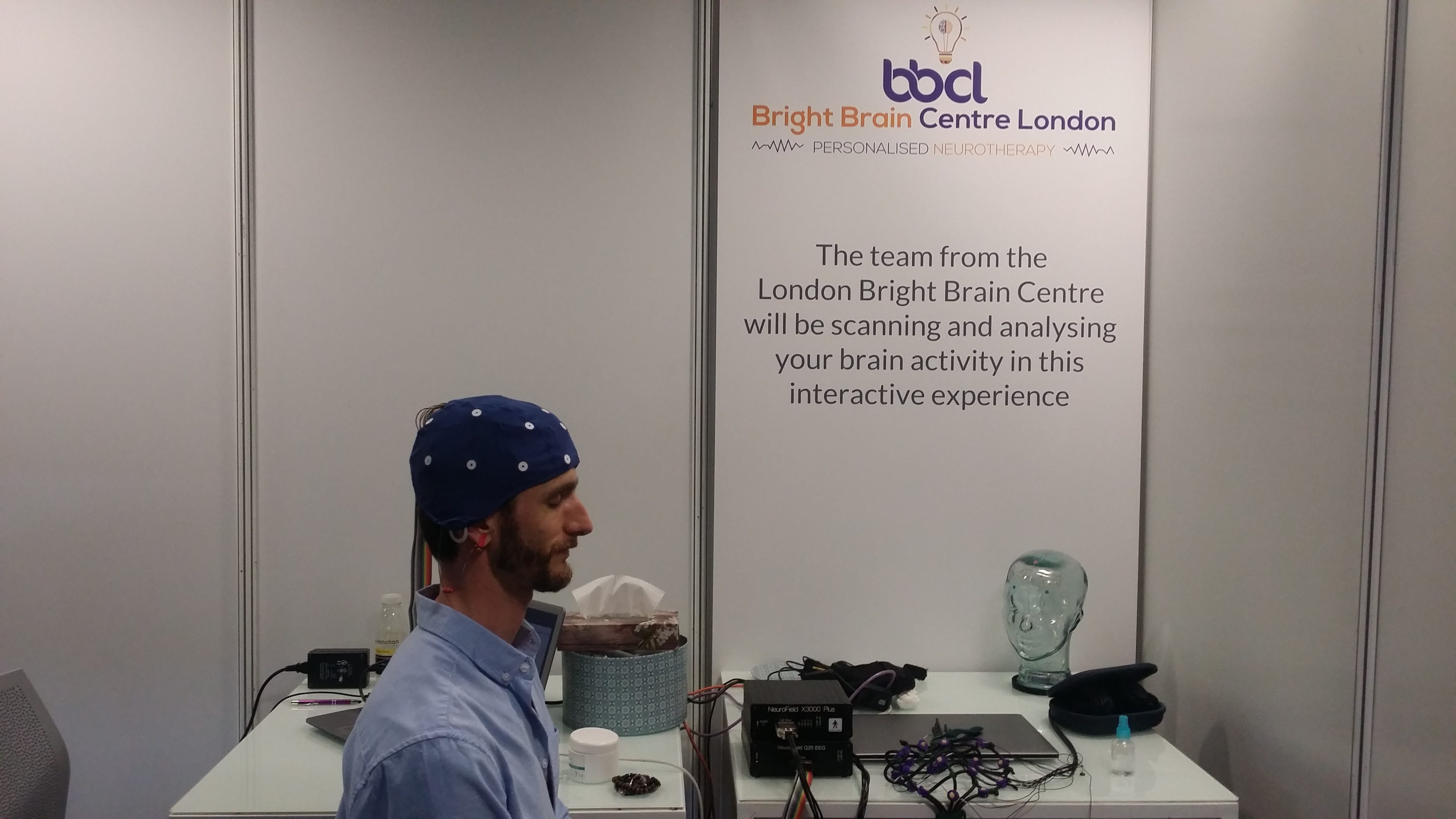
STEP1– Getting Your EEG Reading: What to Expect
When you come in for your 19-channel EEG-recordings with our saline-based free cap, here’s what you can expect:
You’ll simply sit comfortably in our chair while we get everything ready. No special preparation needed on your part! We’ll quickly measure your head to make sure we’re using the right size cap for you. Then we’ll gently place the cap on your head, making sure it’s centered and comfortable.
The cap uses what’s called the “10-20 system” – an internationally recognized method for placing electrodes on your scalp. The name comes from how the electrodes are positioned at points that are either 10% or 20% of the total distance between key landmarks on your head. This standardized approach ensures we capture brain activity from all the important regions. Each electrode has a specific name (like F3, Cz, or O1) that tells us exactly which part of your brain we’re monitoring.
The whole process feels similar to putting on a swimming cap, but much more comfortable.
Preparing the Electrodes AND Checking the Signals
This is where our saline-based system really shines. Instead of using sticky gels or scrubbing your scalp, we simply:
- Part your hair slightly at each electrode spot
- Add a bit of harmless saline solution (just salt water)
- Gently twist each electrode to ensure good contact
You’ll feel a slight coolness from the saline, but no discomfort at all.
Once the cap is on, we’ll connect it to our recording system and check that all electrodes are making good contact. Our screen will show green lights when everything’s perfect. If any spots need a touch more saline, we’ll quickly add it.
During Your Session
Just relax! We’ll have you sit comfortably for a few minutes before starting the actual recording. Throughout your session, we might occasionally add a drop of saline if needed to keep signals clear.
As a part of your brain assessment we typically conduct two resting state EEG recordings to capture the “default” mode of your brain without external distractions. In some cases, we may investigate the speed, accuracy and activation patterns of information processing in your brain, while you perform a computerised neuro-psychological task.
You will be given opportunities to have short breaks, if you feel you need some.
When We’re Done
Removing the cap takes just seconds – we’ll gently lift it off from back to front. We’ll offer you a towel to dry any remaining saline (though there’s usually very little). Your hair might be slightly damp in spots, but the saline dries quickly and leaves no residue.
The entire process typically takes just 30-40 minutes from start to finish, for resting state EEG data collection only. Our clients are often surprised by how quick, comfortable and hassle-free the whole experience is!
If you went on to complete the EEG on task, this would add perhaps another 40 minutes to youre assessment time.
STEP2- LOOKING AT THE DATA
EEG captures your neural communication as it happens- within milliseconds- revealing the dynamic conversation happening in your brain right now. What you’re witnessing is the electrical activity of your mind in its natural state- voltage fluctuations that represent thousands of neurons firing in coordinated patterns. These oscillations aren’t merely abstract data; they’re the fundamental mechanism through which your brain processes information, regulates attention, and generates consciousness itself.
The recording shows the continuous ebb and flow of different frequency bands- from the slower alpha waves that emerge during relaxed alertness to the rapid beta oscillations that underlie active cognition. Each pattern carries specific functional significance, reflecting different aspects of neural processing across distributed networks.
During assessment, we analyze these signals from multiple cortical regions to create a comprehensive map of your brain’s electrical activity. This objective neurophysiological profile reveals both your brain’s intrinsic strengths and areas where communication efficiency might be enhanced.
This precise window into neural function forms the scientific foundation of our neurotherapy approach. By understanding the unique electrical language of your brain, we can design interventions that effectively optimize communication between neural networks, potentially improving cognitive performance, emotional regulation, and overall brain health.
EEG SIGNAL SPECTRAL DECOMPOSITION- 2-D BRAIN MAPS
The Fast Fourier Transform (FFT) provides a quantitative decomposition of the EEG signal into constituent frequency components, enabling precise measurement of spectral power distribution. The power distribution is then “mapped” onto a 2-D surface “heat” maps of the head. Frequently these heat maps represent comparisson with various normative EEG-databases to reveal significant deviations from the normal range.
A Three-Dimensional View: Looking Deeper
Through Low Resolution Electromagnetic Tomography (LORETA), we can transform two-dimensional readings into a three-dimensional map of your brain activity—allowing us to visualize which deeper brain structures might be involved in your unique patterns.
This advanced technique estimates neuronal activity in subcortical regions based on scalp-recorded signals, providing spatial insights that standard EEG cannot achieve. The video demonstrates how LORETA reveals dynamic activity across multiple frequency bands in real-time, with colored intensity maps indicating significant activations compared to normative values.
This enhanced spatial precision is particularly valuable because it helps us understand not just what’s happening but where it’s happening with anatomical specificity. When you describe feeling anxious or having difficulty concentrating, we can often observe corresponding activity in regions like the anterior cingulate cortex, prefrontal areas, or limbic structures that govern these functions.
By identifying specific neural generators of dysregulated activity, LORETA enables us to target neurotherapy interventions with greater precision. Executive function challenges might correlate with underactivation in the prefrontal cortex, while emotional dysregulation often involves hyperactivity in limbic structures like the amygdala.
This three-dimensional mapping bridges the gap between surface EEG and functional neuroimaging, giving us deeper insights that lead to more precisely targeted neurotherapy protocols addressing the underlying neurophysiological origins of your symptoms.
STEP3- BRINGING IT ALL TOGETHER INTO YOUR NEURO-PROGRAMME
Based on these findings, and considering your goals, history, individual learning style and preferences, we develop your personalized brain optimisation program.
This tailored approach combines suitable bio-neurofeedback training sessions with selected non-invasive stimulation methods, addressing the specific neural patterns identified in your assessment, in conjunction with the subjectively uncomfortable patterns you identified as goals to improve through neurotherapy.
THE TRUE VALUE OF YOUR BRAIN ASSESSMENT
The real value of brain mapping lies in its ability to provide objective information about subjective experiences. When you’ve felt that “something’s not quite right” or wondered why certain challenges persist despite your best efforts, brain mapping can offer a physiological perspective that’s often missing from traditional approaches. For many of my clients, seeing their brain patterns brings a profound sense of validation. It’s not “all in your head” in the dismissive sense—it’s literally in your brain, visible and measurable.